Memorable Microanatomy: The Four Basic Tissue Types (Studying Histology Part 2)
In Part 1, we talked about why histology feels confusing at first: students often jump into organ systems before they understand the basic visual language of tissues. The key idea was simple: before trying to identify kidney, liver, lung, thyroid, or intestine, you need to recognize the basic patterns that appear again and again on slides.
That is where the four basic tissue types come in.
Almost every histology slide you will see is built from some combination of four tissue “families”: epithelial tissue, connective tissue, muscle tissue, nervous tissue, and cartilage/bone (specialized connective tissue)
Once you can recognize these families, organ histology becomes less like memorizing random pink and purple blobs and more like reading a visual map.
Think of the body like a building. Epithelium is the wallpaper, tile, and lining. Connective tissue is the glue, padding, wiring support, and scaffolding. Muscle is the moving machinery. Nervous tissue is the communication system.
Your goal is not just to memorize definitions. Your goal is to look at a slide and ask:
What tissue family am I looking at? What pattern proves it? What could I confuse it with?
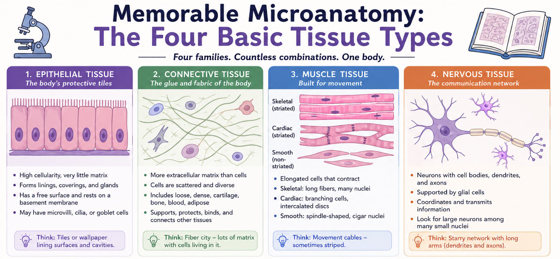
Epithelial Tissue: The Body’s Protective Tiles
Epithelial tissue is usually the easiest tissue to understand conceptually, but it can still be tricky on slides.
Epithelium forms linings, coverings, and glands. It lines the skin surface, digestive tract, respiratory tract, urinary tract, ducts, glands, and many internal spaces. If the tissue is facing a free surface, a lumen, or the outside world, epithelium is probably nearby.
A simple way to imagine epithelium is to think of tiles on a floor or wallpaper on a wall. The cells are packed tightly together, with very little space between them. This is different from connective tissue, where cells are more spread out in a background of matrix and fibers.
When looking for epithelium, focus on four things:
Cell shape
Are the cells flat, cube-shaped, or tall?Number of layers
Is there one layer or many layers?Free surface
Does one side face a lumen or external surface?Special surface features
Do you see cilia, microvilli, keratin, or goblet cells?
For example, in the intestine, epithelial cells form a lining specialized for absorption. In the skin, epithelium forms a protective barrier. In glands, epithelial cells specialize in secretion.
A useful slide clue: epithelial cells often look crowded and organized, almost like rows of students sitting close together in a lecture hall.
Clinical connection: many cancers are called carcinomas because they arise from epithelial tissue. That is one reason epithelial tissue matters so much beyond the microscope.
Quick memory image:
Epithelium = protective tiles lining a surface.
Connective Tissue: More Than Body Glue
Connective tissue is the tissue that students often underestimate. It seems vague at first because it includes many different things: loose connective tissue, dense connective tissue, cartilage, bone, adipose tissue, and even blood.
The key idea is this: connective tissue usually has fewer cells and more extracellular matrix.
That matrix may contain collagen fibers, elastic fibers, ground substance, minerals, or fluid. So instead of seeing tightly packed sheets of cells, you often see cells scattered in a background that may look pink, pale, fibrous, glassy, or spacious depending on the tissue type.
Think of connective tissue as the fabric, glue, scaffolding, and packing material of the body.
It supports epithelium. It surrounds organs. It forms tendons and ligaments. It cushions structures. It provides pathways for blood vessels and nerves. It stores fat. It forms cartilage and bone.
A common place to notice connective tissue is under epithelium. For example, in many organs, you may see a dark, organized epithelial layer facing a lumen, and underneath it, a lighter region with scattered cells and fibers. That supportive layer is often connective tissue.
Important examples include:
Loose connective tissue: more open space, scattered fibers, many cell types.
Dense connective tissue: thick collagen bundles, fewer visible cells.
Cartilage: cells in lacunae surrounded by firm matrix.
Bone: mineralized matrix with osteocytes in lacunae.
Blood: cells suspended in fluid matrix.
A useful slide clue: if there is a lot of “stuff between the cells,” think connective tissue.
This is also where many students confuse collagen with smooth muscle. Collagen often looks wavy and fibrous, while smooth muscle tends to look more cellular and organized into bundles with cigar-shaped nuclei.
Quick memory image:
Connective tissue = fiber city with cells living far apart.
Muscle Tissue: Spot the Stripes
Muscle tissue is built for contraction. On slides, muscle usually appears as elongated cells or fibers arranged in bundles, sheets, or layers.
There are three major types:
Skeletal muscle
Cardiac muscle
Smooth muscle
The first big question is: Do you see striations?
Striations are stripe-like patterns caused by the organization of contractile proteins. Skeletal muscle and cardiac muscle are striated. Smooth muscle is not.
Skeletal Muscle
Skeletal muscle fibers are long, cylindrical, and usually very large. They are striated and often have multiple nuclei, usually located near the edges of the fibers.
On a slide, skeletal muscle can look like a bundle of long pink cables with zebra-like stripes.
Memory image: skeletal muscle = long striped ropes.
Cardiac Muscle
Cardiac muscle is also striated, but it looks different from skeletal muscle. Cardiac cells are shorter, branching, and connected to each other. You may see intercalated discs, which are dark lines between cardiac muscle cells.
Cardiac muscle has a more connected, branching appearance because the heart needs coordinated contraction.
Memory image: cardiac muscle = branching striped network.
Smooth Muscle
Smooth muscle has no visible striations. The cells are spindle-shaped, meaning they are thicker in the middle and tapered at both ends. The nuclei are often described as cigar-shaped.
Smooth muscle is found in places like blood vessels, the gastrointestinal tract, airways, uterus, and bladder. It often appears as pink bundles or layers of elongated cells.
Memory image: smooth muscle = pink waves with cigar nuclei.
A useful trick: when you see pink tissue, do not immediately call it muscle. Ask whether the nuclei and organization fit muscle. Smooth muscle can be confused with dense connective tissue, but smooth muscle usually looks more cellular and has more organized, elongated nuclei.
Quick memory image:
Muscle tissue = movement fibers, sometimes with stripes.
Nervous Tissue: Starry Patterns and Long Arms
Nervous tissue is specialized for communication. It includes neurons, which transmit signals, and glial cells, which support, protect, nourish, and maintain the nervous system.
On slides, nervous tissue can look very different depending on whether you are viewing the brain, spinal cord, peripheral nerves, or ganglia. But the general idea is that nervous tissue is made for sending information across distances.
A neuron has a cell body, nucleus, and long processes such as axons and dendrites. These processes can make neurons look like trees, stars, or cells with long arms.
One helpful image is a starry night: large neuron cell bodies scattered among many smaller glial nuclei.
In nervous tissue, pay attention to size differences. Neurons are often larger, with more obvious cell bodies and nuclei. Glial cells are usually much smaller and more numerous.
In peripheral nerves, you may not see the full neuron cell body. Instead, you may see bundles of axons, often appearing as circular or wavy profiles depending on the section.
A common mistake is expecting every nervous tissue slide to show a perfect textbook neuron with beautiful branches. Real slides are messier. Sometimes you recognize nervous tissue not because it looks dramatic, but because you see large neuron cell bodies mixed with many small supporting cells.
Quick memory image:
Nervous tissue = communication wires and star-shaped signal cells.
Key Takeaways
The four basic tissue types are not just another list to memorize. They are the foundation for understanding almost every organ slide you will see later.
Remember these quick rules:
Epithelium usually forms a lining, surface, or gland. Look for packed cells and a free surface.
Connective tissue has more extracellular matrix and fibers. Look for space between cells.
Muscle tissue has elongated contractile cells. Look for fibers, bundles, and striations.
Nervous tissue is built for communication. Look for neurons, glial cells, and processes.
The goal is not perfection on the first try. The goal is to train your eyes so you understand why you are missing questions on exams. Each time you review a histology slide, ask what pattern you saw, what feature proved the answer, and what look-alike led you toward the wrong choice.
Once you can recognize these four tissue families, histology becomes much less random. You are no longer just memorizing pink and purple shapes, you are learning how to prevent missed questions by reading the body’s microscopic architecture with a clearer, more organized approach.